The Radiology of Warfare
Many papers on the current conflict and on military radiology were published in the BJR in the 1940s. The editorial of November 1941 considered the radiation protection issues involved in the manipulation of fractures and removal of foreign bodies under X-ray control (BJR 1944; 14(167): 343-343).
There were many papers related to war injuries. CJ Hodson gave an account of chest injuries in October 1944 (Hodson BJR 1944; 17(202): 296-299). Lesion associated with severe blows on the chest and with close-up explosions, such as the detonation of cartridges carried in the patient's clothing, occurring without penetrating). In June 1945 a revolver bullet wound at close range (Hodson BJR 1945; 18(176): 176-180).

X-rays showing small entry bullet wound in second left intercostal. Source: Hodson BJR 1945; 18(176): 176-180
L G Blair wrote on thoraco-abdominal injuries in August 1945 (Blair BJR 1945; 18(212): 258-262)
Brigadier D B McGrigor (who came out of retirement for the duration of the war and was Consulting Radiologist to the Army) discussed the training of the military radiologist in 1945 (McGrigor BJR 1945; 18(207): 92-94) and with Lieutenant Colonel Eric Samuel (who was the commanding Officer of the Army X-ray School) wrote an instructive series on the radiology of war injuries discussing abdominal injuries, urinary tract injuries (McGrigor BJR 1945; 18(208): 121-126), thoracic injuries (McGrigor BJR 1945; 18(209) 133-145), head and neck injuries (McGrigor BJR 1945; 18(211): 221-228), orbit and eyeball (BJR 18, 284-290), and jaw and face (McGrigor BJR 1950; 23(276): 685-696).

Image source: McGrigor BJR 1945; 18(208): 121-126
Rowan Williams, from St Mary’s Hospital in Paddington, wrote about the design of diagnostic radiology departments in September 1945 for his BIR Presidential Address (Williams BJR 1945; 18(213): 267-277) and his views are an interesting reflection on the technology and attitudes of the 1940s. The departmental plans are valuable and instructive and reflect the current technology.
 Radiology Department Plan Image Source: Williams BJR 1945; 18(213): 267-277
Neuroradiology
In the 1940s the pioneer neuroradiologist A Schüller was working at the Nuffield Institute for Medical Research in Oxford. In April 1940 he wrote an interesting study of the sub-arachnoid cisterns and their demonstration using a positive contrast agent (Schuller BJR 1940; 13(148): 127-129).
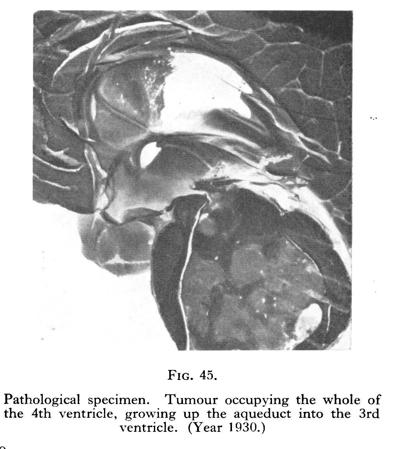
Erik Lysholm from Stockholm gave the Mackenzie Davidson Memorial Lecture for 1946 and spoke about sub-tentorial tumours (Lysholm BJR 1946; 19(227): 437-452).

Image source: Lysholm BJR 1946; 19(227): 437-452
E Lindgren from Stockholm wrote an interesting account of direct puncture percutaneous cerebral angiography in August 1947 (Lindgren BJR 1947; 20(236) 326–331) and the technique was well described. Encephalography and angiography became standard techniques for the investigation of intracranial pathology.
Image source: Lindgren BJR 1947; 20(236) 326–331
James Brailsford wrote a detailed study of cerebral cysticercosis appearing in the March 1941 BJR (Brailsford BJR 1941; 14(159): 79-93).
Obstetrical radiology
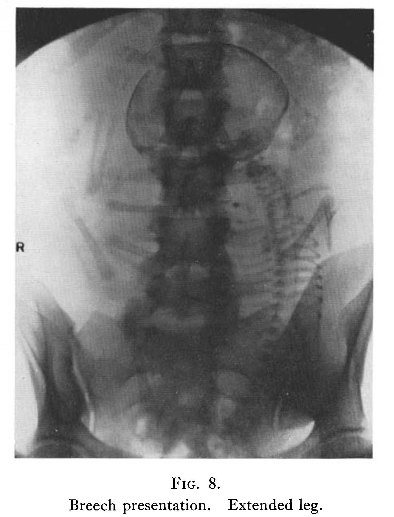
In 1940 the Mackenzie Davidson Memorial Lecture was given by L A Rowden on the subject of maternal mortality and the use of radiology (Rowden BJR 1940; 13(150): 185-192). This was an important topic. In the early 1940s there were over 3000 deaths in childbirth every year, often due to mechanical problems during delivery. Abnormalities of the bony pelvis were far more common then than now and it was important to discover these prior to labour. In 1931 L A Rowden (Rowden BJR 1931; 4(45): 432-439) had demonstrated a simple and effective technique for X-ray pelvimetry.
Image source: Rowden BJR 1940; 13(150): 185-192
Cine-radiology
In 1940 A E Barclay was working at the Nuffield Institute for Medical Research in Oxford and with K J Franklin and M M L Pritchard wrote an interesting paper describing their apparatus for “X-ray cinematography” (Barclay,Franklin and Pritchard BJR 1940; 13(151): 227-234).
 Their apparatus is fully described and illustrated. In March 1942 they described the mechanism of closure of the ductus venosus (Barclay,Franklin and Pritchard BJR 1942; 15(171): 66-71) in an elegant experimental study carried out in the difficult wartime conditions. Further papers in this area of research were to appear (Barclay,Franklin and Pritchard BJR 1942; 15(177): 249-256). The Faculty of Radiologists met in Oxford in 1943 and Barclay spoke on “Radiology – Empiricism or Science?” This thoughtful assessment of the role of the diagnostic radiologist is worth reading. Barclay put a strong case for a scientific approach to radiology and described the great opportunities for radiology in the future.
Image source: Barclay,Franklin and Pritchard BJR 1940; 13(151): 227-234
Contrast media

The use of the new ionic intravascular contrast media was not without risk and in August 1940 F H Kemp describes venous thrombosis following the injection of Uroselectan B (Kemp BJR 1940; 13(152): 291-292).
One use of contrast media is the evaluation of sinus tracts, fistulae and cavities and H Courtney Gage and E Rohan Williams from St. Mary’s Hospital wrote a classic study of this often neglected aspect of radiology in January 1943 (Gage and Williams BJR 1943; 16(181): 8-21). It is still relevant today.
Image source: Gage and Williams BJR 1943; 16(181): 8-21
Skeletal radiology
James Brailsford wrote a detailed account of neuropathic bone changes in October 1941 (Brailsford BJR 1941; 14(166): 320-328) and of osteogenesis imperfecta in May 1943 (Brailsford BJR 1941; 16(185): 129-136). He wrote an interesting account of bone tumours in April 1947 (Brailsford BJR 1947; 20(232): 129-144). Brailsford had an unrivalled experience of skeletal radiology and had followed his patients for many years.
Brailsford gave the Mackenzie Davidson Memorial Lecture in August 1945 and gave his views on the teaching of radiology (Brailsford BJR 1945; 18(212): 249-257) and his views are still relevant today, particularly his thoughts about the dangers of radiological interpretation without appropriate knowledge. In 1946 Brailsford gave a lecture on Wilhelm Conrad Röntgen (Brailsford BJR 1946; 19(227): 453-461) and reviewed his life and contributions in a very readable account and more on his life in a biography
Image source: Brailsford BJR 1947; 20(232): 129-144)
Gastrointestinal tract
Conventional barium examination of the upper and lower gastrointestinal tracts were well developed by the 1940s. Endoscopy, however, was still very primitive and an interesting paper from October 1941 by S Whatley Davidson, a radiologist from Newcastle –on-Tyne, and J Dudfield Rose, who was described as a gastroscopist, is fascinating (Davidson and Dudfield BJR 1941; 14(166): 307-319).  Their subject is the cooperation between the radiologist and endoscopist and is beautifully illustrated by black-and-white barium meal radiographs and colour illustrations of the stomach. The endoscope was rigid in these days before the fibre-optic endoscope. The use of the rigid gastroscope was not without risk as was recorded in a case report by J Duncan White from the Hammersmith Hospital in November 1941 (Duncan BJR 1941; 14(167): 364-365).
Source: Davidson and Dudfield BJR 1941; 14(166): 307-319
Chest radiology
Peter Kerley was Physician to the X-ray Department of Westminster Hospital and Radiologist to the Royal Chest Hospital in June 1942 and wrote a most interesting paper on erythyma nodosum (Kerley BJR 1942; 15 (174): 155-165).  He gives an interesting discussion of sarcoidosis and the radiographs shown are elegant.
Image source: (Kerley BJR 1942; 15 (174): 155-165
In December 1942 he reviewed the techniques needed in mass miniature radiography of the chest (Kerley BJR 1942; 15(180): 346-347) and in 1945 with Kathleen C Clark, P D’Arcy Hart and Brian C Thompson he co-wrote the Medical Research Council Special Report #251 on Mass Miniature Radiography of civilians (HMSO 1945) (Clark, Hart and Thompson BJR 1945; 18(214): 326).
The apparatus needed for mass chest radiography was reviewed by Anthony Nemet in July 1948 (Nemet BJR 1948; 21(247): 319-329).
In July 1943 George Simon from St. Bartholomew’s Hospital reviewed the early changes of pulmonary tuberculosis (Simon BJR 1943: 16(187): 217-219).  Tuberculosis was common in the 1940s and Simon reports an incidence of TB in 14% of patients referred by their general medical practitioners for the exclusion of the disease.
Image source: Simon BJR 1943: 16(187): 217-219
Thomas Lodge from Sheffield was interested in the anatomy of the chest. He published his classic study of the anatomy of the pulmonary vessels in January (Lodge BJR 1946; 19(217): 1-13) and February (Lodge BJR 1946; 19(218): 77-87) 1946. The article is still useful and added considerably to our knowledge of pulmonary anatomy in health and disease. The articles are recommended.
The techniques for bronchography were described by Anthony Vickers from Worcester in March (Vickers BJR 1949; 22(255): 137-151) and April (BVickers BJR 1949; 22(256): 224-233).
Image source: Vickers BJR 1949; 22(255): 137-151
Angiocardiography
The radiology of the heart developed towards the end of the 1940s and KD Keele from Ashford demonstrated an early angiocardiogram in congenital heart disease in August 1948
(Keele BJR 1948; 21(248): 380-393) 
Image source: Keele BJR 1948; 21(248): 380-393
and J A Brocklebank described the technique (Brocklebank BJR 1948; 21(248): 393-397). The conclusion was that the best film interpretation took place when the radiologist and physician reviewed the images together. Israel Steinberg and Charles T Dotter used angiography to distinguish mediastinal tumours from aneurysm in October 1949 (Steinberg and Dotter BJR 1949; 22(262): 567-572).
There were many variations of apparatus made for angiography and angiocardiography and M McGregor who worked with the cardiologist John McMichael at Hammersmith Hospital described one such table in August 1949 (McGregor BJR 1949; 22(260): 459-461) and J D F Williams from Coventry described another in November 1949 (Williams BJR 1949; 22(263): 666-668). 
X-ray table of angiocardiography Source: Williams BJR 1949; 22(263): 666-668
Nuclear medicine
In November 1948 George Ansell and Joseph Rotblat from Liverpool demonstrated a retro-sternal thyroid using radioactive iodine and external counting (Ansell and Rotblat BJR 1948; 1(251): 552-558)  is a classic and often referred to paper.
Geiger Counter
Image source: Ansell and Rotblat BJR 1948; 1(251): 552-558
The end-window Geiger counter is illustrated and their complete apparatus is described. The intra-thoracic thyroid tissue was outlined accurately. They also indicated that the radio-iodine could also be used for the treatment of hyperthyroidism. This paper was the first of many to appear in the BJR of the field that would become known as Nuclear Medicine.