Radiology in the NHS
The 1960s was a time of many changes, both social and scientific. The NHS had now been established for some years and Sir George Godber from the Ministry of Health reviewed the position of radiology in BJR August 1963 (Godber BJR 1963; 36(428): 545-548). Radiology was becoming increasingly complex and expensive. Sir Harry Platt spoke of “Medicine at the crossroads” in 1965 at the middle of the decade (Platt BJR 1965; 38(452): 561-566) and there were strong feelings of uncertainty and frustration in both the medical profession and the country.
Knowledge was expanding in all areas. Bernard Lovell described the radio telescope in his Silvanus Thompson Memorial Lecture of 1961 (Lovell BJR 1962; 35(409): 1-4) and Robert Platt described advances in genetics in March 1960 (Platt BJR 1960; 33(387): 134-137). FG Spear in his Presidential Address of 1962 reviewed the current state of the Institute (Spear BJR 1962; 35(410): 77-89). There is an interesting account of the origins of the BIR device of a spiked sun and the similarities to the solar disc symbol of Aten.
There was a more critical approach to clinical practice and in March 1960 James Bull and others from the National Hospital in Queen Square, London, looked at inter-observer variability in image interpretation (Bull, Couch, Joyce et all BJR 1960; 33(387):165-170).
New techniques were appearing to replace traditional radiological procedures. GD Scarrow from Liverpool described the new Olympus fibre-optic endoscope and camera in January 1967 (Scarrow BJR 1967; 40(469): 23-29) with coloured endoscopic photographs as illustrations. He commentated on the often poor correlation between the barium meal and subsequent surgical results.

Image source: Scarrow BJR 1967; 40(469): 23-29
A number of deaths were recorded:
James Frederick Brailsford
James F Brailsford died in 1961 (Grout BJR 1961; 34(400): 269). Brailsford’s contributions to skeletal radiology were massive and his book on the radiology of bones and joints was highly influential and is available in the historical book collection of the Institute.
James Brailsford was active in radiology at an interesting time in its history. The period of the pioneers had passed and radiology was developing as a specialty. By the 1930s there had developed a systematic interpretation of the shadows cast on the X-ray film. Not only was radiology developing but there was now specialisation within radiology. The early radiologists were involved in both diagnosis and radiotherapy, however by the 1930s radiology was dividing into those who primarily specialised in radiotherapy and those who spent their time in diagnosis.
It was at this time that James Brailsford’s great book on ‘The Radiology of Bones and Joints’ was published. The book was issued in 1934 and Brailsford dedicated it to Sir Robert Jones, the pioneer orthopaedic surgeon who had done so much to develop orthopaedic surgery as a science. In the preface to his book, Brailsford describes how radiography had extended our knowledge of the growth, development and structure of the bones and joints and both health and disease. Brailsford also realised that the technique of radiography required a specialist for both performing examinations and for image interpretation. It was therefore important that the radiologist took an active part in research, diagnosis, treatment and prognosis. If the radiologist was not involved in these areas then Brailsford saw that the radiologist was no more than a qualified technician. Brailsford became deeply involved in defining the professional role and responsibilities of the radiologist.

Image source: Grout BJR 1961; 34(400): 269
Ernest Rock Carling (1877-1960)
Ernest Rock Carling was a surgeon to Westminster Hospital who became very interested in radiation hazards. His obituary was written by WV Mayneord (Mayneord BJR 1960; 33(393): 593).

Thomas James Case (1882-1960)
James Case was a pioneer American radiologist and his obituary was written by James Brailsford (Brailsford BJR 1960; 33(394): 617).
Kathleen (Kitty) Clara Clark (1896-1968)
Kathleen (Kitty) C Clark died in 1968 (BJR 1968; 41(492): 948). Miss ‘Kitty’ Clark completed her training course at Guy’s Hospital in 1921 and passed the first ever qualifying examination ever held by the Society of Radiographers. The Society of Radiographers had been set up in 1920 and letters had been written from the new society to assistants in the various X-ray departments inviting applications for membership. Those who been in active practice for over of 10 years were automatically given membership without examination. All other applicants had to take a new examination.
The first regular batch of students was entered for examination in January 1922. There were 45 students of which twenty passed and were duly awarded the certificate of the Society (the MSR). Miss ‘Kitty’ Clark initially worked in at the Princess Mary’s Hospital Margate before moving to the Royal Northern Hospital in London.
She was aware of the lack of adequate training for radiographers, and so she founded a School of Radiography at the Royal Northern Hospital which became a model for schools elsewhere.
In 1935 she became co-founder and Principal of the Ilford Radiographic Department at Tavistock House, where she was involved in instruction and research into radiography and medical photography. Under her guidance the department developed a world-wide reputation.
She was President of the Society of Radiographers from 1935 to 1937 (and also the first woman President). She was the first President to wear the President’s chain of office, it having been presented by her predecessor Dr Leo Rowden.
The first edition of her classic book ‘Positioning in Radiography’ (Heinemann Medical Books) was published in 1939 (BJR 1939; 12(136): 252). The book became the standard work of reference for radiographers and has been through many editions. Her slide collection has been preserved and is available in the library of the British Institute of Radiology as the ‘K C Clark Slide Library’. Although primarily of historical interest, the slide collection is still a useful teaching resource and may be copied.
‘Positioning in Radiography’ is a very interesting book for several reasons. Firstly, it standardized the radiographic projections and so similar projections were made in all hospitals. Kitty Clark was keen to standardize both positioning and exposure. Secondly, the book is very artistic. The illustrations do not come across as cold and entirely objective scientific images. It is therefore not surprising to read that the artist Francis Bacon acknowledged ‘Positioning in Radiography’ as a crucial source and it was his favorite medical textbook. Lawrence Gowling indicated that Bacon repeatedly borrowed from the photographs in the book for his work. The images of the body that Francis Bacon made have an almost radiographic quality and there is the impression that multiple layers of the body are seen at the same time and that one is not just looking at the skin surface.
She was awarded the MBE in 1945 for her services to radiography, particularly for mass miniature radiography of the chest. Her book: Mass Miniature Radiography of Civilians (MRC special report series No. 251) and written jointly with P D’Arcy Hart, Peter Kerley & Brian Thompson appeared in 1945 (D’Arcy Hart et al. BJR 1945; 18(214): 326).
She was committed to fostering co-operation and contact between radiographers throughout the world and was a driving spirit behind the formation of the ISRRT. The ISRRT is an organisation composed of seventy-one national radiographic societies from sixty-eight countries representing more than 200,000 radiographers and radiological technologists.
She remained as principal at Ilford until 1958 and acted as Consultant in Radiography until 1964.
John Cockcroft
Sir John Cockcroft died in 1967 (Allibone BJR 1967; 40(479): 872-873) and made many contributions to the nuclear industry and its applications to science and medicine.

Image source: Allibone BJR 1967; 40(479): 872-873
Neville Samuel Finzi (1881-1968)
Neville Finzi died in 1968 (IGW BJR 1968; 41(487): 552). Finzi was a pioneer and made many contributions to radiotherapy and is remembered in the Finzi Lecture of the Radiology Section of the Royal Society of Medicine. His book “Radium Therapeutics” was published in 1913 and gives an account of his treatment of carcinoma of the oesophagus using intra-luminal Radium.

Image source: IGW BJR 1968; 41(487): 552
Felix Fleischner
Felix Fleischner died in 1969 and his obituary was written by Robert Steiner (Steiner BJR 1969; 42(504): 947). Fleischner was a major figure in the developments of chest radiology and is remembered in the Fleischner Society
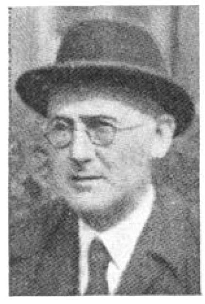
Louis Harold Gray
The death of LH Gray in 1965 marked the end of an era in radiobiological research. His obituary was written by Jack Boag (Boag BJR 1965; 38(453): 706-707). EL Powers gave an American view of Gray in October 1965 (Powers BJR 1965; 38(454): 804-805). Louis Harold Gray gave his BIR Presidential Address in 1950 and spoke on non-medical aspects of medical radiology (Gray BJR 1950; 23(275): 627-633). Gray had entered medical physics in 1933 and he discusses his experiences. His thoughts are interesting. For example, damage due to a single ionizing particle will be in direct proportion to the dose and if the damage cannot be reversed by the cell then the damage will be independent of the dose-rate. For a given dose there will be a certain probability of producing cell damage however small the dose given and so the old concept of a tolerance dose-rate which presupposed a threshold rate of exposure below which no damage would occur. It follows that a permissible level of exposure can only logically be set at such a level that there is no significant increase over and above damage arising from natural causes.
In 1952 Gray gave the second Douglas Lea Memorial Lecture and gave a magnificent account of the action of radiation of the living cell (Gray BJR 1952; 25(293): 235-244). Gray discussed Lea’s work and then gave an account of his own understanding of this topic. In 1953 he delivered the Silvanus Thompson Memorial Lecture and spoke on the initiation and development of cellular damage by ionizing radiation (Gray BJR 1953; 26(312): 609-618).
The Gray (Gy) is the derived SI unit for absorbed dose, specific energy and kerma (kinetic energy in matter). This SI unit was named after Gray. 1 Gray is the dose of energy that is absorbed by a homogeneously distributed material with a mass of 1 kilogram when it is exposed to ionising radiation bearing 1 joule of energy (1 Gy = 1 J/kg).

Image source: Boag BJR 1965; 38(453): 706-707
Sidney Russ (1879-1963)
Sidney Russ died in 1963 and had started work in a hospital in 1910 being appointed the physicist in 1913. He was probably the first hospital physicist in the world. (JER BJR 1963; 36(429): 702). He was a supporter of the BIR through his career, and he joined the Röntgen Society in 1910. Further appreciation of Sidney Russ appeared in November 1963 (Andrews et al. BJR 1963; 36(431): 862-83).

Image source: JER BJR 1963; 36(429): 702
Rolf Maximilian Sievert (6 May 1896 – 3 October 1966)
Rolf Sievert, who died in 1966, was a Swedish medical physicist who worked on the measurement of radiation dosage and studied the biological effects of radiation (FWS BJR 1967; 40(470): 142). In 1979 the SI unit for ionizing radiation dose equivalent was named in his honour as the Sievert (Sv). The Sievert (symbol: Sv) is the SI derived unit of dose equivalent and reflects the biological effects of radiation in contradistinction to the physical aspects, which is the absorbed dose and measured in Grays (Boag BJR 1965; 38(453): 706-707).

Image source FWS BJR 1967; 40(470): 142
Rohan Williams
Rohan Williams was only 56 when he died in 1961 and he was still President of the Faculty of Radiologists (DWS BJR 1963; 36(424): 300).