Ian Kelsey Fry
In the 1980s the technology that had been started in the 1970s was gradually becoming more widely available. However, such technology did not come without a cost. The theme of “Who needs high technology?” was chosen by Ian Kelsey Fry for his presidential address of 1983 (Kelsey Fry BJR 1984; 57(681): 765-772). The 1980s was a time of pressures on resources and the use of expensive medical equipment had to be justified. Ian Kelsey Fry concluded by saying that our patients need high technology and that it is our job to ensure that we evaluate the technology properly and that, whenever possible, patients are provided with the level of technology appropriate to their clinical needs. Many traditional techniques passed out of use in the 1980s, being replaced by better and more efficacious modalities. In 1970 42% of antenatal patients had an X-ray examination and this had reduced to 3% by 1980. The changing role of radiology in obstetrics was reviewed by A Gordon and others (Gordon, Pinchen, Walker and Tudor BJR 1984; 57(682): 891-893) in October 1984.

Image source: Kelsey Fry BJR 1984; 57(681): 765-772
CT scanning
As the 1980s progressed the papers describing the use of CT became increasing sophisticated and complex. Many of the techniques described at that time are now part of our routine daily practice. GAS Lloyd, PD Phelps and GH Du Boulay from London wrote a beautifully illustrated paper on high-resolution CT of the petrous bone in July 1980 (Lloyd, Phelps and du Boulay BJR 1980; 53(631): 631-641). A further paper on CT air meatography followed in January 1982 (Phelps and Lloyd BJR 1982; 55(649): 19-22) and on high resolution CT of the petrous bone in July 1982 (Lloyd and Phelps BJR 1982; 55(655): 483-491).
The paper by AP Nisbet and others from London in June 1983 on clinical indications for the optimal use of the radionuclide brain scan (RBS) in an age of CT is interesting (Nisbet, Ratcliffe, Ellam, Rankin and Maisey BJR 1983; 56(666): 377-381). Whilst the authors indicate that as a physiological technique the RBS is complimentary to the CT brain scan the technique was fighting a losing battle as the number of CT scanners slowly increased. In a similar manner CT was to fight a losing battle as MRI improved in quality and availability although the introduction of CT spiral volumetric scanning in the 1990s reversed the trend.
CT continued to improve in quality with technical developments such as 3-D images shown by JE Gillespie and Ian Isherwood from Manchester in March 1986 (Gillespie and Isherwood BJR 1986; 59(699): 289-292), A Gholkar and Ian Isherwood in March 1988 (Gholkar and Isherwood BJR 1988; 61(723): 258-261), and A Gholkar and others in November 1988 (Gholkar, Gillespie, Hart, Mott and Isherwood BJR 1988; 61(732): 1095-1099). They used 3-dimensional reformats and whilst the images are elegant it was following the development of helical scanning that the technique came into its own.

Image source: Gholkar, Gillespie, Hart, Mott and Isherwood BJR 1988; 61(732): 1095-1099
Nuclear medicine
MV Merrick and others from Edinburgh recorded their experience in the detection of pyelonephritic scars in children by radioisotope imaging (Merrick, Uttley and Wild BJR 1980; 53(630): 544-556) with correlation of nuclear medicine and urography.
In September 1980 NW Nicholson and others from Manchester wrote about HIDA scanning in gall-bladder disease (Nicholson, Hastings, Testa and Torrance BJR 1980; 53(633): 878-882).
Both of the above two tests are still in active clinical use today. It was not obvious at the time which techniques will survive and which will pass away. HIDA and DMSA survive and the use of thallium 201 for the diagnosis of cerebral metastases (Ancri and Basset BJR 1980; 53(629);443-453) and selenomethionine liver scanning for the diagnosis of hepatoma (Coakley and Wraight BJR 1980; 53(630): 538-543) pass away.
In February 1981 C Kellershohn from France gave his Mackenzie Davidson Memorial Lecture on the use of positron-emitting radionuclides (PET scanning) (Kellershohn BJR 1981 54,(638): 91-102) and was a masterly account of the current experience. In November 1982 K Schelstraete and others from Belgium studied the uptake of 13N-ammonia by human tumours again using PET (Schelstraete, Simons, Deman, Vermeulen and Siegers, Vandecasteele, Goethals and De Schryver BJR 1982; 55(659): 797-804).
AM Peters and others from the Hammersmith wrote a paper for the November 1982 journal describing some of their experience in indium labelled white cells in the diagnosis of infection (Peters, Karimjee, Saverymuttu and Lavender BJR 1982; 55(659): 827-832).
Pulmonary embolism remained a problem and in January 1986 JP Finn and others from Hammersmith Hospital assessed the then new DTPA aerosol system for lung ventilation scanning (Finn, Myers, Nair and Lavender BJR 1986; 58(697): 19-24). This technique became a standard.
Towards the end of the decade RT Ott from the Royal Marsden Hospital reviewed nuclear medicine in his Mayneord Lecture of 1988 (Ott BJR 1989; 62(737): 421-432) . The then state of nuclear medicine was examined followed by a detailed look at positron emission tomography (PET scanning). PET scanning has become of increasing usefulness now combined with CT scanning (PET-CT) and in the lecture Ott was calling for regional cyclotron facilities and lower-cost positron camera systems.
Radiographic contrast media
In the 1980s the conventional high osmolar ionic contrast agents were gradually replaced by the modern non-ionic and low osmolar agents. RG Grainger from Sheffield wrote about the omolality and described the new contrast media in August 1980 (Grainger BJR 1980; 53(632): 739-746). In 1981 RG Grainger gave his Mackenzie Davidson Memorial Lecture on intravascular contrast media (Grainger BJR 1982; 55(649): 1-18). This lecture is required reading for anyone at all interested in contrast media. Of particular interest is the story of Moses Swick. The development of modern contrast media has transformed medical imaging and made many other developments possible. Peter Dawson and Michael Howell from London reviewed the new non-ionic dimmers in October 1986 (Dawson and Howell BJR 1986; 59(706): 987-991).
Peter Dawson reviewed contrast agent nephrotoxicity in February 1985 (Dawson BJR 1985; 58(686): 121-124) and this remains a clinical concern.

Image source: Dawson BJR 1985; 58(686): 121-124
Ultrasound
Peter Wells from Bristol was the BIR president in 1986 and chose as the topic of his presidential Address “The Prudent Use of Diagnostic Ultrasound” (Wells BJR 1986; 59(708): 1143-1151). Caution needs to be taken with the introduction of any new technique. In April 1989 Peter Wells reviewed Doppler ultrasound (Wells BJR 1989; 62(737): 399-420) with a review of the principles.
Ultrasound was increasingly used in pregnancy as the 1980s progressed and P Farrant from Northwick Park described the early ultrasound diagnosis of fetal bladder neck obstruction (Farrant BJR 1980; 53(629): 506-508). In August 1981 with H Meire she published a study establishing the normal fetal limb lengths in pregnancy (Farrant and Meire BJR 1981; 54(644): 660-664).
In pregnancy both the changes in the fetus and the mother can be examined. In May 1985 KA Cietak and JR Newton from Birmingham Maternity Hospital elegantly defined the quantitative changes in the maternal kidney as pregnancy progresses (Cietak and Newton BJR 1985; 58(689): 405-413). Brian J Cremin from Cape Town gave a beautiful review of real time abdominal ultrasound in children in September 1985 (Cremin BJR 1985; 58(693): 859-868) and PB Guyer and KC Dewbury from Southampton produced excellent images of benign breast disease (Guyer and Dewbury BJR 1988; 61(725):374-378).
As the 1980s progressed there was increasing use of imaging guided biopsy procedures. In October 1982 G Montali and others described their experience of real-time biopsy of liver lesions using a needle inserted into the central canal of a real-time linear-array ultrasound probe (Montali, Solbiati, Croce, Ierace and Ravetto BJR 1982; 55(658): 717-723). In September 1985 Per G Lindgren described excision biopsy of the spleen under ultrasound guidance using a spring loaded handle containing a Tru-Cut® type core biopsy needle (Lindgren, Hagberg, Eriksson, Glimelius, Magnusson and Sundström 1985; 58(693): 853-857).

Image source: Lindgren, Hagberg, Eriksson, Glimelius, Magnusson and Sundström 1985; 58(693): 853-857
MRI
In October 1981 the first biomedical images to be produced using the new high speed echo-planar imaging technique were published by RJ Ordidge, P Mansfield and RE Coupland from Nottingham (Ordidge, Mansfield and Coupland BJR 1981; 54(646): 850-855). The paper was illustrated in colour and clear anatomical detail was shown. The possibility for imaging moving objects and flow was discussed.
By June 1983 JM Kean and others from Nottingham showed what strides had been made in a short time by publishing a clinical paper on MRI of the knee (Keen, Worthington, Preston, Roebuck, McKim-Thomas, Hawkes, Holland and Moore BJR1983; 56(666): 355-364). Although by modern standards the images appear primitive there is excellent visualisation of anatomical detail and a clear indication of what a powerful tool for musculo-skeletal imaging MRI was to become. So as an example look at the April 1993 paper by CW Heron from St George’s Hospital (Heron BJR 1993; 66(784): 292-302) on MRI of the knee only 10 years later.
The MRI equipment at Nottingham was home-build and improvements allowed them to produce snap-shot images. This was explored in September 1988 (Howseman, Stehling, Chapman,Coxon, Turner, Ordidge, Cawley, Glover, Mansfield and Coupland BJR 1988; 61(729): 822-828) looking at abdominal imaging. Despite the major technical obstacles the image quality gradually improved.
The group at Nottingham were interested in moving images shown on NMR (MRI). RG Ordidge, P Mansfield and others described such real time movies of the heart in the October 1982 BJR (Ordidge, Mansfield, Doyle and Coupland BJR 1982; 55(658): 729-733) depicting frames from a movie loop of the chest of a live rabbit. In December 1983 M Doyle and others from Nottingham were describing dynamic NMR (MRI) cardiac imaging in a piglet (Doyle, Rzezdian, Mansfield and Coupland BJR 1983; 56(672): 925-930).
Continuing the use of MRI in the cardiovascular system, HG Bogren from the National Heart and Chest Hospitals in London produced elegant images of velocity mapping in aortic dissection (Bogren, Underwood, Firmin, Mohiaddin, Klipstein, Rees and Longmore BJR 1988; 61(726): 456-462).
MRI has a major role in the CNS. M Graif and RE Steiner from Hammersmith Hospital gave an elegant account of contrast enhanced MRI of brain tumours in September 1986 (Graif and Steiner BJR 1986; 59(705): 865-873) and RR Mawhinney and others from Nottingham gave an equally elegant account of the cerebello-pontine angle in October 1986 (Mawhinney, Buckley and Worthington BJR 1986; 59(706): 961-969).
The surface coils that are used in MRI imaging are illustrated in a paper by Heather Deans and others from Aberdeen (Deans, Redpath, Smith, Parrek and Forrester BJR 1988; 61(728): 665-672) published in the August 1988 BJR. The group used surface coils over the eyes producing excellent images of orbital anatomy and pathology.
Contrast media were used in MRI as well as in conventional imaging. In September 1988 JP Stack and others from Manchester reviewed the use of gadolinium-DTPA in imaging for acoustic neuroma (Stack, Ramsden, Atoun, Lye, Isherwood and Jenkins BJR 1988; 61(729): 800-805) producing elegant images and demonstrated that scanning times could be reduced considerably.
As well as imaging the MR spectroscopy is important. Rolf D Oberhaensli and others including George Radda from the John Radcliffe Hospital in Oxford (Oberhaensli, Galloway, Hilton-Jones, Bore, Styles, Rajagopalan, Taylor and Radda BJR 1987; 60(712):367-373) reviewed the study of human organs by phosphorus -31 topical magnetic resonance spectroscopy. Spectra for brain and liver were obtained and the clinical uses were reviewed.
In October 1988 towards the end of the decade Graham Bydder from the NMR Unit at the Hammersmith Hospital gave a very nice review of the then state of play of MRI (Bydder BJR 1988; 61(730): 889-897). The first clinical MRI had been produced in the UK in Nottingham and Aberdeen in 1980 and by 1988 there were only 24 clinical MRI systems in the UK compared to 150 CT scanners.

Image source: Bydder BJR 1988; 61(730): 889-897)
Mammography
Many techniques have been used to image the body. Xeroradiography enhances edges and the images were illustrated in colour in April 1980 (Keddy and Brebner BJR 1980; 53(628): 325-330).
In June 1982 EJ Roebuck (Roebuck BJR 1982; 55(654): 387-398) from Nottingham published a detailed paper describing the importance of mammographic parenchymal patterns. In September 1982 G Hermann and others from New York’s Mount Sinai Hospital discussed the radiological presentation of non-palpable breast tumours (Hermann, Janus, Mendelson and Brady BJR 1982; 55(657): 623-628).
Towards the end of the decade in 1988 that AMP Forrest gave his Mackenzie Davidson Memorial Lecture on the topic of screening for breast cancer in the UK (Forrest BJR 1988; 62(740): 695-704). In 1987 a national mammographic programme for screening for breast cancer was initiated. The lecture reviews the natural history of breast cancer and then looks at the benefits of screening. The UK national screening programme is described and discussed fully.
Lymphography
Lymphography was used to diagnose both lymphatic disease and malignancy. In an elegant study J McIvor and others from Charing Cross Hospital in London described in February 1980 the appearances of metastatic disease in lymphography performed in patients with prostatic cancer (McIvor, Massouh, Backhouse and MacRae BJR 1980; 53(626): 74-80).
Angiography/interventional radiology
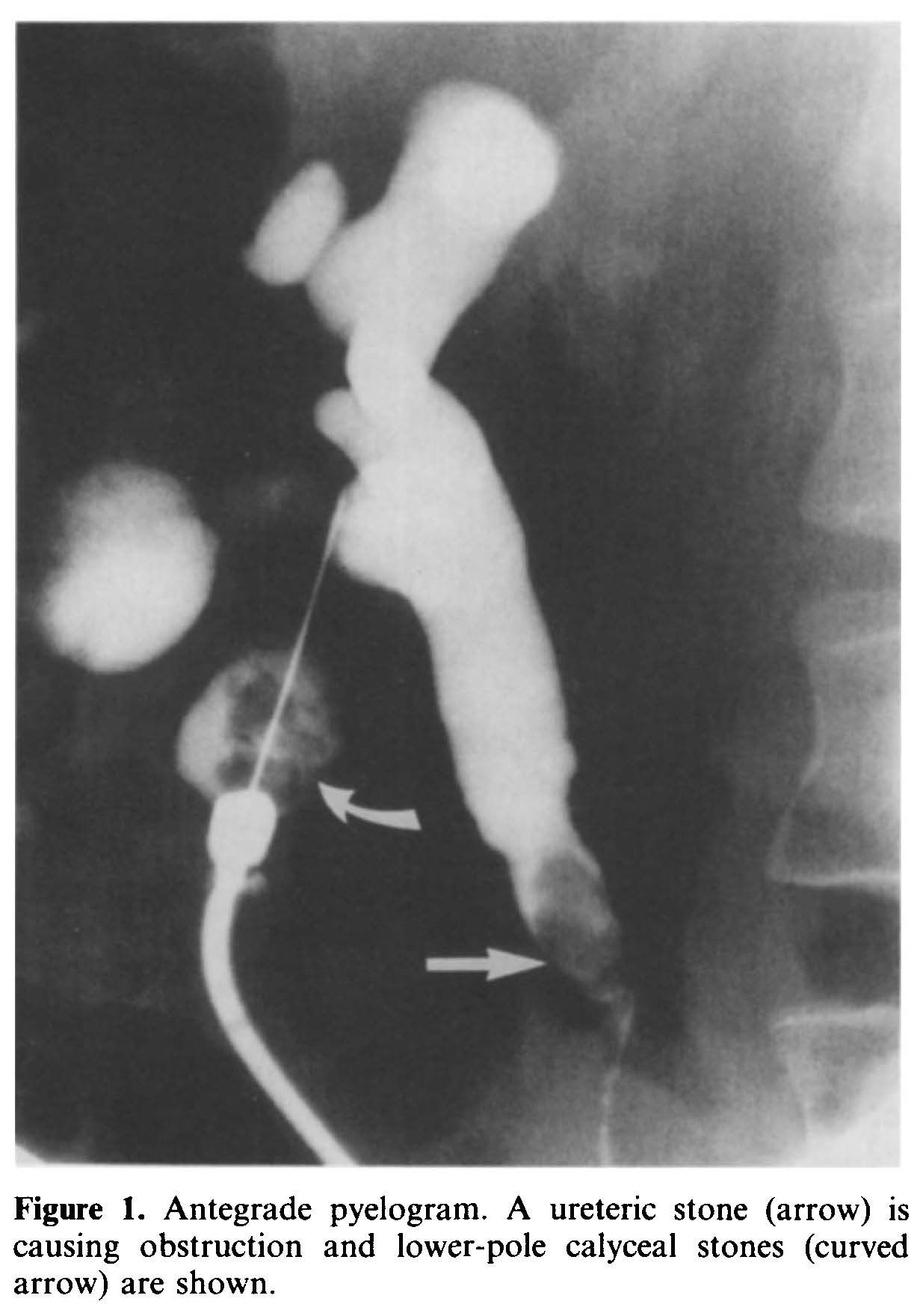
In a very interesting paper in August 1982 by JW Ludwig and others on digital video subtraction angiography (Ludwig, Verhoeven and Engels BJR 1982; 55(656): 545-553). The apparatus is described and the principles are illustrated. The varieties of images that could be obtained are beautifully depicted. It was the development of the rapid digital subtraction techniques and rapid digital angiography that greatly facilitated the development of the new sub-speciality of interventional radiology. The applications of the new discipline of interventional radiology to the renal tract were beautifully illustrated by D Rickards and SN Jones from the Middlesex Hospital in July 1989 (Rickards and Jones BJR 1989; 62(739): 573-581).

Image source: Rickards and Jones BJR 1989; 62(739): 573-581