36 Portland Place home of BIR from 1980s
The major event of the 1980s was the opening of the new home for the institute at 36 Portland Place. Her Majesty the Queen, as patron, visited us on February 11th 1982. A great deal of work had taken place to enable the move to occur and the Institute remains grateful to Sir Jan Lewando and Mr Kevin Hughes who were joint Chairmen of the Appeal Committee and to Prof. George Du Boulay who was the Appeal Coordinator. Her Majesty the Queen had graciously granted her Patronage to the Institute in 1979 and this is duly recorded in the pages of the journal (Bewley and Roylance BJR 1980; 53(629): 395-397).
In 1986 the BIR celebrated 90 years of X-rays and held a joint meeting with the NRPB and the BAAS at the Royal Society on November 8th. The abstracts were published in the BJR (BJR 1986; 59(703): 717-722) and are all worth looking at.
The presidential address that William M Ross had given at the 1979 BIR Annual Congress was reprinted in the February 1980 BJR (Ross BJR 1980; 53(626): 55-62). The paper is well worth reading and summarises the development of the Institute and its future prospects. His view of the Institute that “the major attraction is the opportunity for members of all crafts and sciences associated with radiology to meet on an equal footing”.
Radiation accident
Sadly the major event involving radiation in the 1980s was the accident at the Chernobyl nuclear power station in the Ukraine on the 26th April 1986 at 01.23 local time. For the 1987 Mayneord Lecture FA Fry reviewed the accident at Chernobyl (Fry BJR 1987; 60(720): 1147-1157) and the paper still makes chilling reading. The BIR held a one-day seminar as part of its 1987 Annual Congress and the papers are all reprinted. CC Lushbaugh and others from Oak Ridge in Tennessee spoke on radiation accident management (Lushbaugh, Fry and Ricks BJR 1987; 60(720): 1159-1163), JC Nénot from France spoke on the establishment of levels for medical intervention (Nénot BJR 1987; 60(720):1163-1169), NM Nadezhina from the USSR spoke on organizing a specialized centre for medical care (Nadezhina BJR 1987; 60(720): 1169-1170), PJ Bourdillon from the DHSS spoke on the role of NHS hospitals in the event of a nuclear accident (Bourdillon BJR 1987; 60(720): 1171-1174), B Edmonson from London spoke on nuclear reactor design in the UK (Edmonson BJR 1987; 60(720): 1174-1177), JK Wright spoke on emergency planning for nuclear incidents (Wright BJR 1987; 60(720):1177-1180), RAF Cox on medical preparedness for nuclear emergencies (Cox BJR 1987; 60(720):1180-1182) and finally Roger H Clarke from the NRPB finished with a talk putting reactor accidents into perspective (Clarke BJR 1987; 60(720):1182-1188).

Image source: Fry BJR 1987; 60(720): 1147-1157
Many of the papers in the early 1980s still describe the use of traditional imaging to investigate patients that today would go straight to cross-sectional imaging (CT and MRI) or ultrasound. This illustrates that CT scanning was still of limited availability in the 1980s and many hospitals could only acquire a scanner after going to public appeal and do the fund raising themselves. Medical imaging technology continued to advance and these advances had significant effects both on clinical practice and on the economics of medicine. These changes were elegantly and well described by Alexander R Margulis and William Shea from California in their Mackenzie Davidson Memorial Lecture of 1986 (Margulis and Shea BJR 1986; 59(700): 309-315). It’s still worth reading as is the Presidential Address of the BIR given by Ian Isherwood (Isherwood BJR 1986; 59(703): 643-652) entitled “The golden age: a shifting spectrum.” Technology is advancing with great speed and as Prof. Isherwood said: “We stand before a 20th century Rosetta stone - let us use our multidisciplinary strengths, embodied in the traditions of the BIR, to ensure that our scientific vocabulary is appropriate for the 21st century.”
The Silvanus Thompson Memorial Lecture for 1981 was given by John Mallard from Aberdeen (Mallard BJR 1981; 54(646): 831-848). John Mallard made major contributions in many areas and the paper gives a very interesting account of the development of the pioneer MRI unit in Aberdeen. His title was “The noes have it! Do they?” The John Mallard Lecture of The Institute of Physics and Engineering in Medicine was established in 2004 and is given annually at the UK Radiological Congress (UKRC) to recognise the contributions of John Mallard in the field of Medical Imaging. John Mallard pioneered many of the technologies including Nuclear Medicine, Magnetic Resonance Imaging and Positron Emission Tomography. Having established his reputation at the Hammersmith Hospital and St Thomas' Hospital Medical School in London, Mallard took up the first Chair in Medical Physics and Bioengineering in Aberdeen, combining an academic role in the University with a high-class comprehensive physics service to the hospital.
Radiation is used for many indications other than the medical and GB Schofield from British Nuclear Fuels gave his Mackenzie Davidson memorial Lecture in 1979 on the subject of biological control in a plutonium facility (Schofield BJR 1980; 53(629): 398-409) concluding that a careful surveillance of the health of plutonium workers gave no cause for concern. Sadly radiation may be used in warfare and March 1983 a BIR Working Party reported on the radiological effects of nuclear war (BJR 1983; 56(663): 147-170) and sadly the report is still relevant and makes salutary reading.

Image source: Schofield BJR 1980; 53(629): 398-409
The issues that were of concern are still with us, such as the effects of delayed reporting of radiographs considered by G de Lacey and others from St George’s Hospital in April 1980 (de Lacey, Barker, Harper and Wignall BJR 1980; 53(628): 304-309).
In 1981 the BIR president David Trapnell gave a masterly account of chest radiology with a review of the development of chest imaging and a discussion of the physiological principles involved in chest imaging (Trapnell BJR 1982; 55(650): 93-107). Also of interest is the Mackenzie Davidson Memorial Lecture of 1982 given by Robert E Steiner (Steiner BJR 1983; 56(662): 75-85) on the topic of ischaemic heart disease. At this time there is depicted the integration of many imaging techniques with angiocardiography, coronary angiography, nuclear medicine, CT scanning and MRI scanning. The sheer variety of techniques available for a single disease would be astonishing to a previous generation. To illustrate this point further, the Mackenzie Davidson Memorial Lecture for 1985 was given by Torgny Greitz (Greitz BJR 1985; 58(696): 1149-1163) on the topic of diagnostic and therapeutic strategies in neuroradiology. The lecture is illustrated by CT scans (for imaging, treatment planning and stereotaxis), “functional” PET scanning and digital angiography. Of particular interest is the integration of images from different modalities.
During the 1980s there was increasing use of computers in both imaging and therapy departments (Morrey, Smith, Belcher et al BJR 1982; 55(652): 283-288). In December 1982 RF Mould gave a detailed account of the use of a computer to provide the Westminster Hospital Cancer Registry (Mould BJR 1982; 55(660): 897-904). The whole topic of digital imaging was reviewed in detail by RL Smathers and WR Brody from Stanford University in California for the April 1985 (Smathers and Brody BJR 1985; 58(688): 285-307).
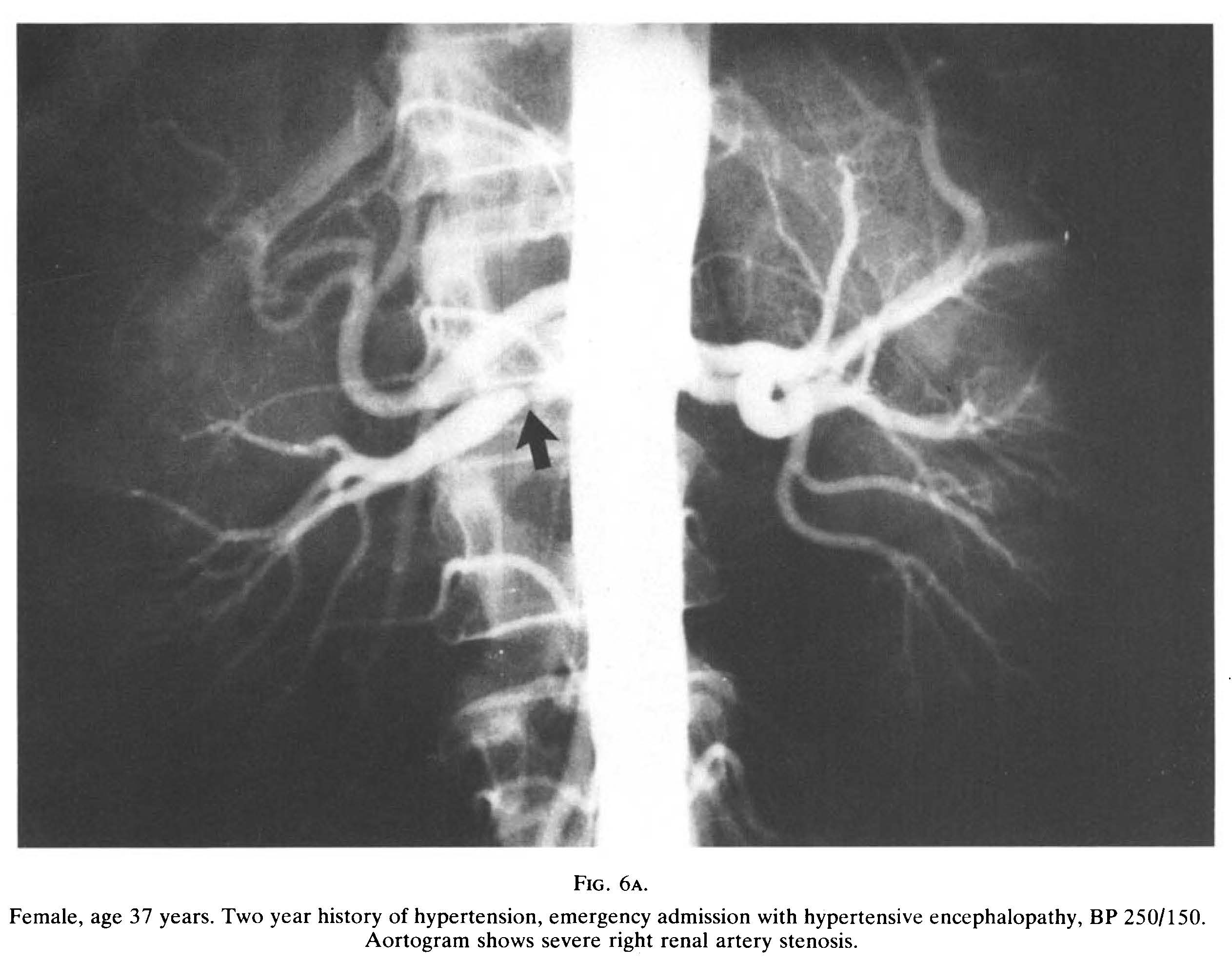
In the 1980s the sub-speciality of interventional radiology was developing rapidly. In May 1982 DC Cumberland described his experience of coronary, renal and peripheral angioplasty (Cumberland BJR 1982; 55(653): 330-337). Cumberland emphasized the need for close co-operation between disciplines for optimal case selection and to deal with any complications. Angioplasty had first been described by CT Dotter and MP Judkins in 1964.

Image source: Cumberland BJR 1982; 55(653): 330-337